Magnetic resonance imaging (MRI)
What is Magnetic Resonance Imaging?
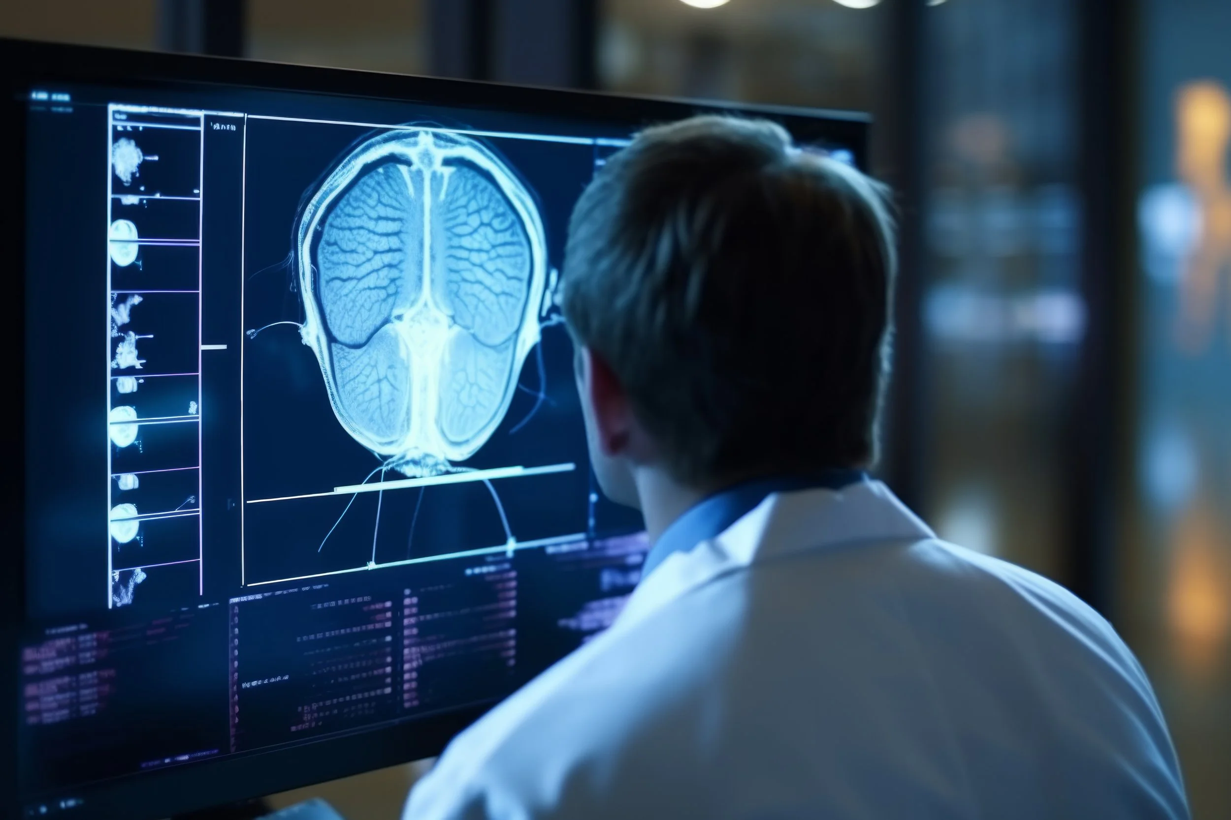
Magnetic resonance imaging (MRI) uses a strong magnet and radio waves to provide clear and detailed diagnostic images of internal body organs and tissues. It is a valuable tool for the diagnosis of a wide range of conditions, including cancer, heart and vascular disease, stroke, and joint and musculoskeletal disorders. MRI allows the evaluation of some body structures that may not be as visible as other diagnostic imaging methods.
WHAT ARE SOME COMMON USES OF MRI?
MRI is frequently used for imaging of the musculoskeletal system. MRI is often used to study the knee, ankle, foot, shoulder, elbow, wrist, and hand. MRI is also a highly accurate method for the evaluation of soft tissue structures, such as tendons and ligaments, which are seen in great detail. Even subtle injuries are easily detected. In addition, MRI is used for the diagnosis of spinal problems, including disc herniation, spinal stenosis, and spinal tumors.
MRI is used for imaging of the heart. MRI of the heart, aorta, coronary arteries, and blood vessels is a tool for diagnosing coronary artery disease and other heart problems. Doctors can examine the size and thickness of the chambers of the heart and determine the extent of damage caused by a heart attack or heart disease. MRI is also used for imaging of cancer and functional disorders. Organs of the chest and abdomen, such as the liver, lungs, kidney, and other abdominal organs, can be examined in great detail with MRI. This aids in the diagnosis and evaluation of tumors and functional disorders. In the early diagnosis of breast cancer, MRI is an alternative to traditional x-ray mammography. Furthermore, because there no radiation exposure is involved, MRI is often used for examination of the male and female reproductive systems.
WHAT IS MR ANGIOGRAPHY (MRA)?
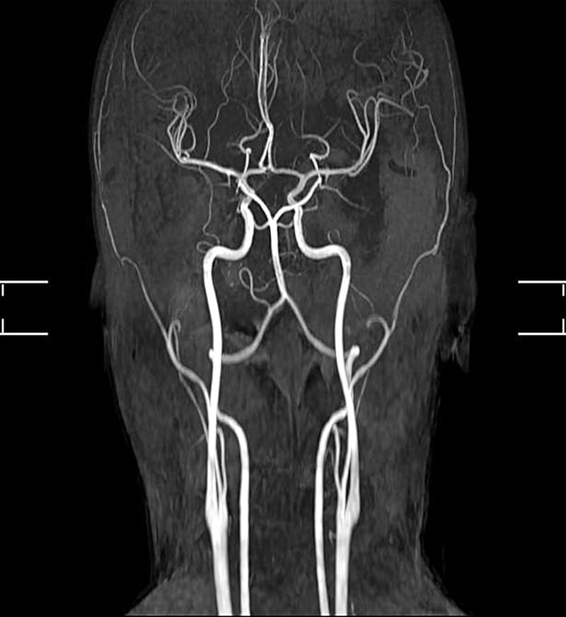
MR angiography (MRA) uses a powerful magnetic field, radio frequency waves, and a computer to evaluate blood vessels and help identify abnormalities or diagnose atherosclerotic (plaque) disease. This exam does not use ionizing radiation and may require an injection of gadolinium, a contrast material less likely to cause an allergic reaction than iodinated contrast material. If needed, the contrast material is usually administered through a small intravenous (IV) catheter placed in a vein in the arm.
WHAT ARE SOME COMMON USES OF MRA?
MRA is used to examine blood vessels in key areas of the body, including the brain, neck, heart, abdomen, pelvis, and extremities (arms, legs, feet, and hands).
HOW SHOULD I PREPARE?
Before your MRI exam, remove all accessories, including hair pins, jewelry, eyeglasses, hearing aids, wigs, and dentures. During the exam, these metal objects may interfere with the magnetic field, affecting the quality of the MRI images taken. Notify your technologist if you:
have any prosthetic joints – hip, knee
have a heart pacemaker (or artificial heart valve), defibrillator, or artificial heart valve
have an intrauterine device (IUD)
have any metal plates, pins, screws, or surgical staples in your body
have neuro-stimulators
have inner ear implants
have tattoos and permanent make-up
have a bullet or shrapnel in your body, or ever worked with metal
are pregnant or suspect you may be pregnant
WHAT SHOULD I EXPECT DURING THIS PROCEDURE?
The exam generally takes 15 to 45 minutes, depending on the number of images needed. However, very detailed studies may take longer. You must lie down on a sliding table and be comfortably positioned. Even though the technologist must leave the room, you can communicate with them anytime using an intercom. You will be asked to remain still during the actual imaging process; however, slight movement is allowed between sequences, which last between 2-15 minutes. Depending on the part of the body being examined, a contrast material may be used to enhance the visibility of specific tissues or blood vessels. A small needle is placed in your arm or hand vein, and a saline solution IV drip will run through the intravenous line to prevent clotting. About two-thirds of the way through the exam, the contrast material is injected.
WHAT WILL I EXPERIENCE DURING THIS PROCEDURE?
MRI is painless. Some claustrophobic patients may experience a “closed-in” feeling. If this is a concern, a sedative is available. Our open, high-field MRI provides a more comfortable, relaxing experience. You will hear loud tapping or thumping during the exam. Earplugs or earphones may be provided to you. You may feel warmth in the area being examined. This is normal. If a contrast injection is needed, there may be some discomfort at the injection site. You may also feel a cool sensation at the site during the injection.


